CIE A Level Biology復習筆記7.1.3 Xylem Vessels Elements

Xylem Vessel Elements: Structure & Function
- The functions of?xylem?tissue in a plant are:
- Vascular tissue that?transports?dissolved minerals and water around the plant
- Structural?support
- Food storage
- Xylem tissue is made up of four cell types that function together:
- Tracheids (long, narrow tapered cells with pits)
- Vessel elements (large with thickened cell walls and no end plates when mature)
- Xylem parenchyma
- Sclerenchyma cells (fibres and sclereids)
- Most of the xylem tissue is made up of?tracheids?and?vessel elements, which are both types of?water-conducting cell
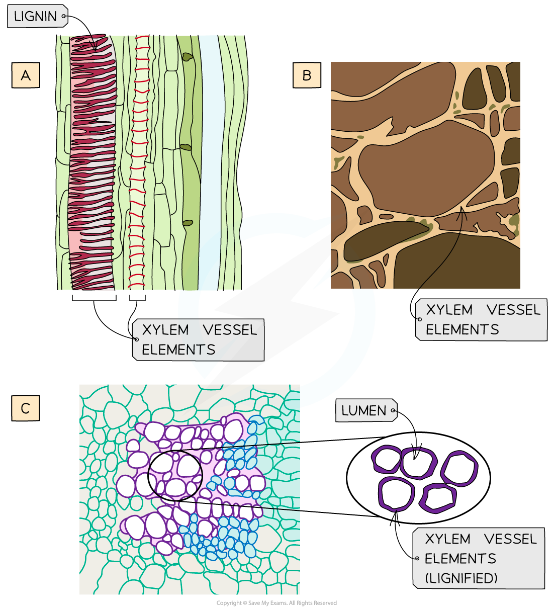
Images of xylem vessel elements, (a) photomicrograph in longitudinal section (lignin is stained red), (b) scanning electron micrograph in transverse section and (c) microscope image in transverse section and drawing (lignin is stained red)
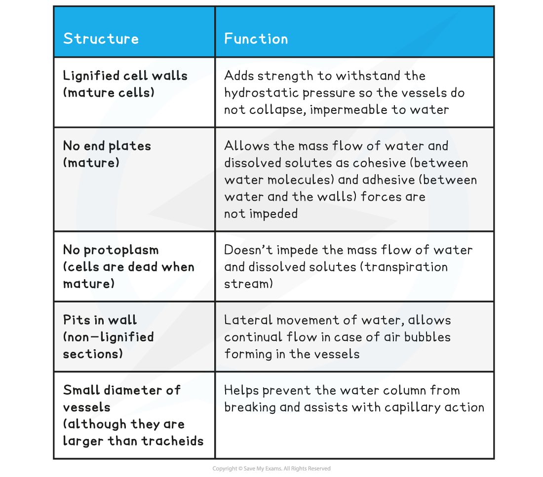
Relating structure & function in xylem vessel elements table
- Also see?Comparison of xylem & phloem tissue table?in Phloem Sieve Tube Elements
Exam Tip
You must be able to recognise the xylem vessel elements in images so look for the thicker cell walls and the larger diameter. You also need to know the difference between xylem and phloem tissue.
轉載自savemyexams
以上就是關于【CIE A Level Biology復習筆記7.1.3 Xylem Vessels Elements】的解答,如需了解學校/賽事/課程動態,可至翰林教育官網獲取更多信息。
往期文章閱讀推薦:
MIT官方發布【2026年夏季推薦閱讀書單】!橫跨科學/人文/經濟...
全網破防!ALevel CIE數學M1疑似錯題?經濟P2難度飆升?5月6日大考考情分析必看!

翰林AMC8視頻課重磅上線!

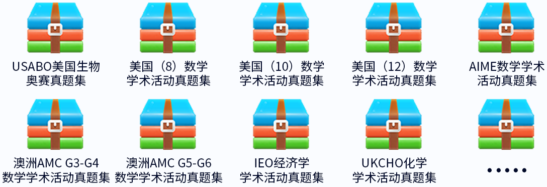
國際競賽真題資源免費領取